

What Is Thermal Imaging?
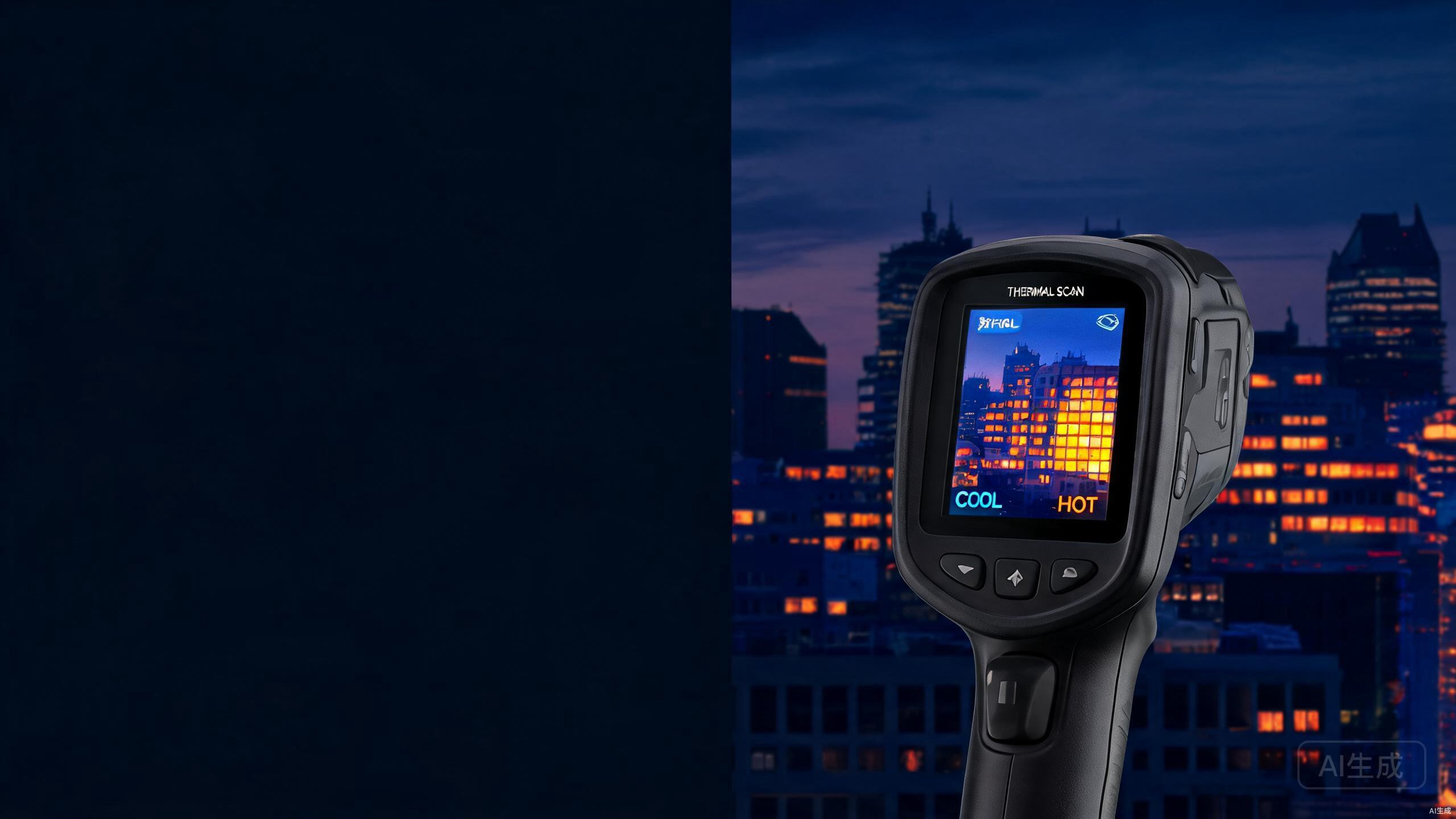
Thermal imaging is a technology that allows us to visualize heat. Instead of capturing visible light like a conventional camera, a thermal imaging device detects infrared radiation—the invisible energy that every object above absolute zero emits. This radiation carries information about the object's temperature, and thermal imaging technology translates that information into images we can see.
Think of it as giving your eyes the ability to perceive temperature differences. A wall with hidden water damage, an electrical panel with an overheating component, or an animal hiding in complete darkness—all of these reveal themselves through their thermal signatures. The core principle is simple: hotter objects emit more infrared energy, and thermal imagers measure that energy to create a visual map of temperature distribution.
This technology does not rely on ambient light. It works equally well in bright daylight, total darkness, fog, or smoke. That independence from visible light conditions is what makes thermal imaging valuable across so many industries.
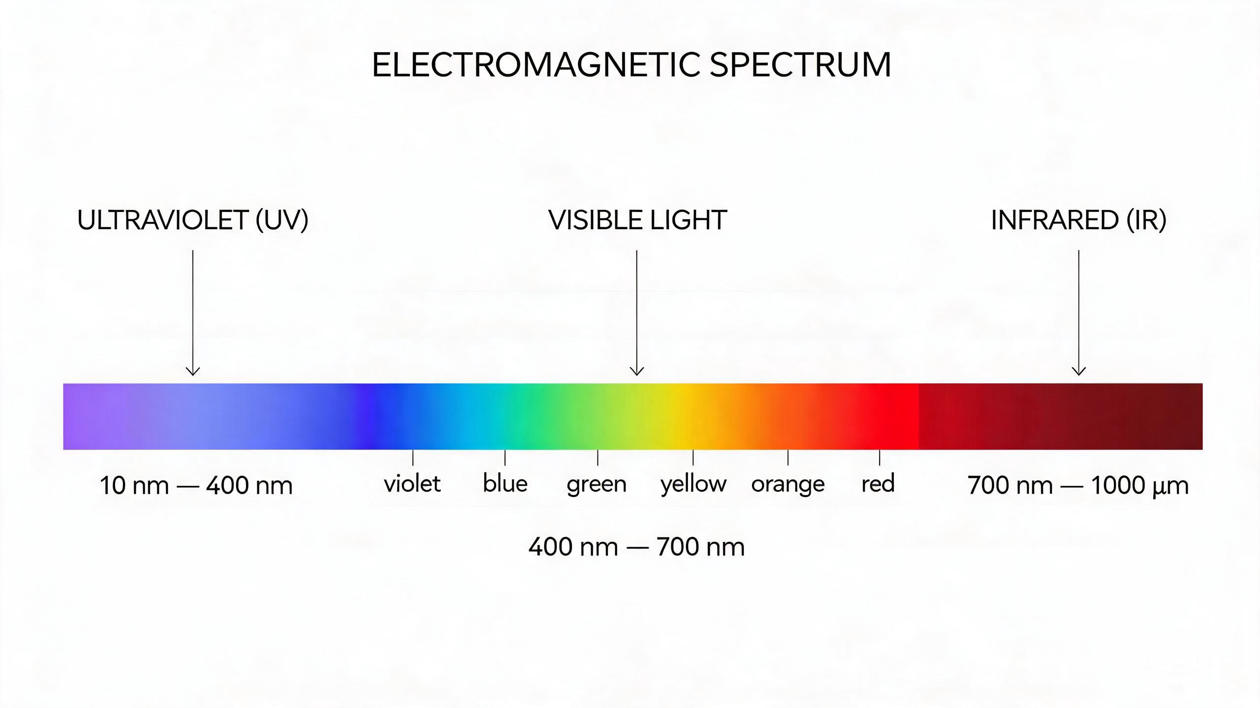
The electromagnetic spectrum showing where infrared radiation sits relative to visible light.
The Science Behind Infrared Radiation
To understand how thermal imaging works, it helps to know where infrared radiation sits in the electromagnetic spectrum. Visible light—the light our eyes can detect—occupies a narrow band between approximately 380 and 700 nanometers in wavelength. Infrared radiation lies just beyond the red end of visible light, with wavelengths ranging from about 700 nanometers to 1 millimeter.
All matter with a temperature above absolute zero (-273.15°C or 0 Kelvin) emits infrared radiation. The amount and wavelength of this radiation depend directly on the object's temperature. This relationship is described by Planck's law of black-body radiation: as temperature increases, the intensity of emitted radiation rises, and the peak wavelength shifts toward shorter (more energetic) wavelengths.
For objects at typical ambient temperatures (roughly -40°C to 100°C), the peak emission falls within the long-wave infrared (LWIR) range, between 8 and 14 micrometers. This is why most thermal imaging systems are designed to detect LWIR radiation. Human skin, for example, at approximately 33°C, emits strongest in this band.
How Thermal Imaging Works: The 4-Step Process
A thermal imaging system converts invisible infrared radiation into a visible image through four sequential stages. Each stage relies on specialized hardware to pass information to the next, forming a complete infrared radiation detection process.
Infrared Lens Captures Radiation
The process begins at the infrared lens. Unlike standard camera lenses made from glass, infrared lenses use specialized materials such as germanium, silicon, or chalcogenide glass. These materials are transparent to infrared wavelengths but opaque to visible light. The lens focuses incoming infrared radiation from the scene onto the detector array behind it. The lens design—whether fixed focal length or zoom—affects the field of view and spatial resolution of the final image.
Detector Converts Radiation to Electrical Signals
Behind the lens sits the infrared detector, the heart of the system. The detector consists of a large array of tiny sensing elements (pixels), typically made from vanadium oxide (VOx) microbolometers or amorphous silicon. When infrared photons strike these elements, they cause a measurable change in electrical resistance or voltage. Each pixel generates an electrical signal proportional to the intensity of infrared radiation it receives. A 640x512 detector, for example, contains 327,680 individual sensing points, each reporting its own temperature reading.
Signal Processing Circuit Amplifies the Signal
The raw electrical signals from the detector are extremely weak. A signal processing circuit amplifies these signals to usable levels and performs initial noise reduction. This stage also applies correction algorithms to compensate for detector non-uniformity—a natural variation in sensitivity across different pixels. Without this correction, the resulting image would contain fixed patterns of noise that obscure the actual thermal scene.
Image Processing Converts Signals to Visual Images
In the final stage, the processed signals feed into an image processing circuit. This unit maps the temperature data to a color or grayscale palette that human eyes can interpret. Common palettes include white hot (hotter objects appear white), black hot (hotter objects appear black), and rainbow or ironbow (using color gradients to represent temperature). The output is a real-time video stream or still image showing the thermal landscape of the observed scene.
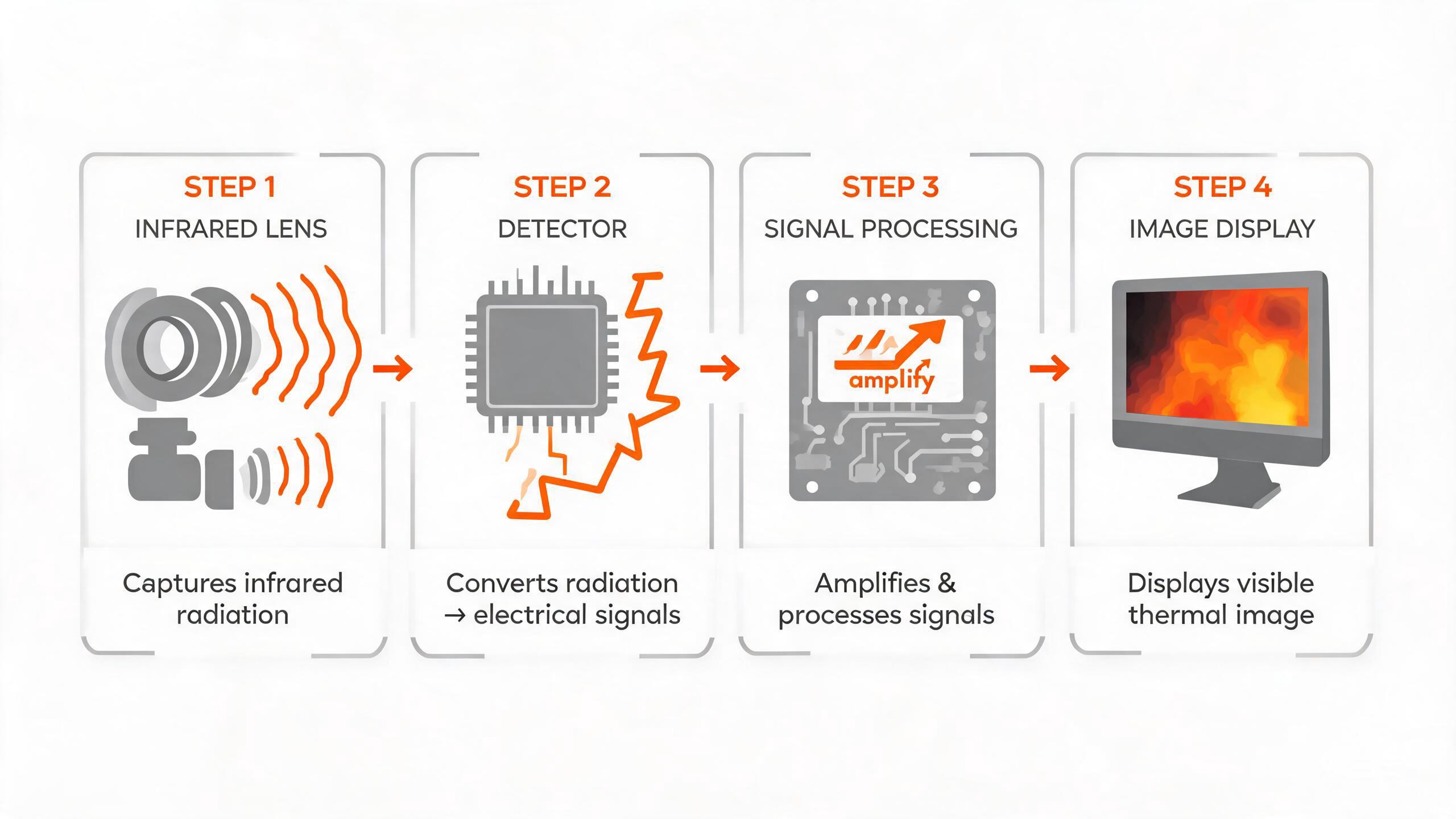
The four-step process of thermal imaging: from infrared capture to visible image output.
Understanding Thermal Imaging Detectors
The detector is the single most important component in a thermal imaging system. Its performance largely determines image quality, sensitivity, and cost. Modern thermal imagers predominantly use uncooled detectors, which operate at ambient temperature without requiring cryogenic cooling.
Uncooled detectors, such as VOx microbolometers, work by measuring temperature changes in the sensing material caused by absorbed infrared radiation. They are compact, energy-efficient, and affordable, making them suitable for handheld cameras, thermal scopes, and integrated modules. Their typical sensitivity, measured as NETD (Noise Equivalent Temperature Difference), ranges from 30mK to 50mK—meaning they can distinguish temperature differences as small as 0.03°C to 0.05°C.
Cooled detectors, in contrast, use cryogenic cooling (often with a Stirling engine) to reduce the detector's operating temperature to around 77 Kelvin (-196°C). This dramatically reduces thermal noise and increases sensitivity, with NETD values often below 20mK. Cooled systems offer superior long-range detection and can resolve finer temperature differences. However, they are larger, heavier, more expensive, and require regular maintenance. They are typically reserved for military, aerospace, and high-end scientific applications.

Comparison of uncooled and cooled thermal imaging detectors and their typical applications.
Types of Thermal Imaging Lenses
The lens in a thermal imaging system does more than focus light. Because infrared radiation behaves differently from visible light, thermal lenses require specialized design and materials. Manufacturers typically offer several thermal imaging lens types to match different operational needs.
| Lens Type | Key Characteristic | Best For |
|---|---|---|
| Fixed-Focus Lens | Maintains focus at a specific distance; simplest and most cost-effective | Fixed-mount surveillance, industrial monitoring with constant target distance |
| Manual-Focus Lens | User rotates focus ring to adjust; compensates for temperature-induced blur | Handheld cameras for building and electrical inspection |
| Motorized-Focus Lens | Internal motor adjusts focus remotely via software | PTZ cameras, unmanned systems, remote monitoring |
| Continuous Zoom Lens | Multiple seamless field-of-view settings; most complex and expensive | High-end security, military platforms, advanced hunting scopes |
Key Applications of Thermal Imaging Technology
Thermal imaging has moved far beyond military origins. Today it serves as a practical tool across civilian industries, each leveraging the ability to see heat signatures invisible to conventional cameras. Here are the thermal imaging applications explained across major sectors.
Medical Diagnosis
Non-invasive visualization of skin temperature to identify inflammation and monitor blood flow.
Building Inspection
Locate insulation gaps, moisture intrusion, and air leaks for improved energy efficiency.
Security & Surveillance
Detect human heat signatures in complete darkness, fog, and smoke for perimeter protection.
Electrical Inspection
Identify overheated components and loose connections before catastrophic failure.
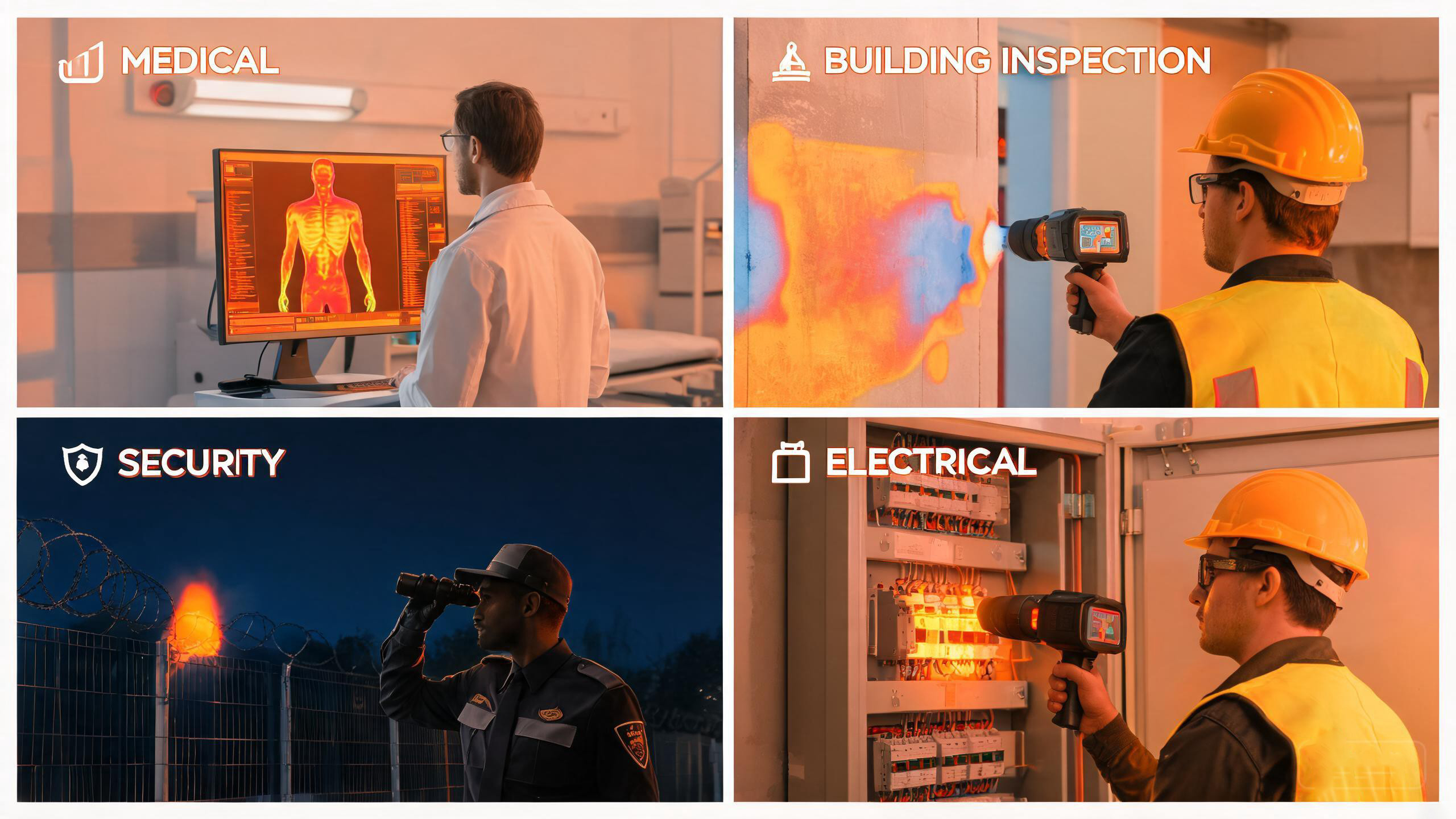
Thermal imaging technology applied across medical, building, security, and electrical industries.
Frequently Asked Questions
A: No. Thermal imaging cannot see through solid walls. It detects surface temperature, not objects behind barriers. However, it can reveal temperature differences on a wall's surface caused by heat transfer from objects or conditions behind it—such as insulation gaps, water damage, or electrical hotspots—without penetrating the material itself.
A: Night vision devices amplify existing visible light or near-infrared light to create an image. They require some ambient light to function and struggle in total darkness or through fog and smoke. Thermal imaging, in contrast, detects long-wave infrared radiation emitted by objects based on their temperature. It works in complete darkness and can see through light fog and smoke, because it does not rely on reflected light.
A: Detection range depends on the detector resolution, lens focal length, and target size. A high-resolution 640x512 detector paired with a long focal length lens can detect a human-sized target at 1,500 to 2,000 meters under ideal conditions. Smaller targets or lower-resolution systems reduce this range. Environmental factors such as humidity, rain, and atmospheric particles also attenuate infrared signals and shorten effective range.
A: Yes. Thermal imaging functions equally well in daylight and darkness because it detects emitted infrared radiation rather than reflected visible light. In fact, bright sunlight can sometimes create challenging thermal conditions where surfaces heat unevenly, but the technology itself is not affected by visible light intensity.
A: Most uncooled thermal cameras designed for general use detect temperatures from approximately -40°C to 550°C. Specialized industrial cameras extend this range to 1,500°C or higher for applications such as furnace monitoring or metal processing. The exact range depends on the detector calibration and the system's intended application.
A: Thermal detectors measure temperature, not color. False color palettes—such as white hot, black hot, ironbow, and rainbow—map temperature values to visible colors or grayscale shades. This makes it easier for human observers to interpret temperature differences at a glance. White hot, for example, assigns white to the hottest temperatures and black to the coldest, a convention that aligns with intuitive understanding.
Explore Thermal Imaging Solutions
Whether you need thermal imaging core modules, cameras, or scopes, Owlshine offers reliable infrared solutions for your application.
Browse Our Products